Traumatic brain injury (TBI) is a major cause of death and disability worldwide, especially in children and young adults. It occurs when the head violently hits an object, or when an object accidentally enters the brain. Currently there is no good way to reverse the brain damage caused by TBI. But doctors can help stabilize the TBI patients and prevent further brain damage. So early detection is critical for the treatment of TBI. Many biomarkers have been associated with the severity of TBI. In 2018, the first blood test was approved by FDA for the evaluation of mild TBI in adult. This test measures two protein biomarkers, UCH-L1 and GFAP, in the blood after brain injury. In 2021, the first rapid handheld TBI blood test has received the clearance from FDA, which also measures UCH-L1 and GFAP in the blood.
TBI Biomarker Panel – Create your custom panel
| Biomarker | GFAP | NEUROFILAMENT | NSE | PGP9.5 | S100 BETA | TAU |
|---|---|---|---|---|---|---|
| Catalog-No. |
TA500338S
30 ul Anti-GFAP mouse monoclonal antibody, clone OTI4D11 (formerly 4D11) |
TA801110S
30 ul NEFL mouse monoclonal antibody,clone OTI3G2 |
UM870177
30 ul NSE(ENO2) mouse monoclonal antibody, clone UMAB287 |
UM870136
30 ul UCHL1 (PGP9.5) mouse monoclonal antibody,clone UMAB244 |
TA807235S
30 ul S100B mouse monoclonal antibody,clone OTI3G6 |
TA809201S
30 ul MAPT mouse monoclonal antibody,clone OTI13B5 |
Click the tabs below for more details:
Ubiquitin C-terminal Hydrolase L1 (UCHL1)
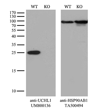
UCHL1 is a neuron-specific enzyme that has been used as a diagnostic biomarker in TBI blood tests. It is highly expressed in brain, testes, and carcinomas of various tissue origins. Many studies have shown a significant negative correlation between UCHL1 levels and the severity of TBI outcome. UCHL1 has shown a better sensitivity than GFAP and S100B as a biomarker to differentiate the TBI patients with normal and abnormal CT head findings. However, UCHL1 alone is not enough to evaluate TBI. In the current FDA approved TBI blood tests, UCHL1 is used together with another important biomarker, GFAP.
Products for UCHL1
Glial Fibrillary Acidic Protein (GFAP)
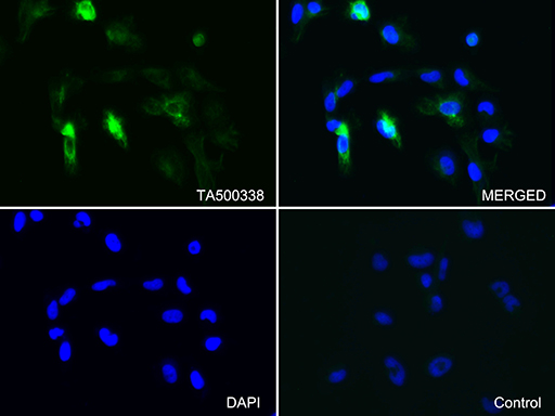
GFAP is a intermediate filament protein found in different cells of the central nervous system (CNS). It is a specific biomarker that can be used for the evaluation of TBI patients. Previous studies have shown GFAP has a good diagnostic accuracy in differentiating mild TBI patients from non-TBI controls. Comparing to S100B and UCHL1, GFAP works better as a predictor for positive CT of the head in TBI patients, even though it may not be a good predictor for the long term outcome. It has been used together with UCHL1 in the current FDA approved TBI blood tests.
Products for GFAP
Neuron Specific Enolase (NSE)
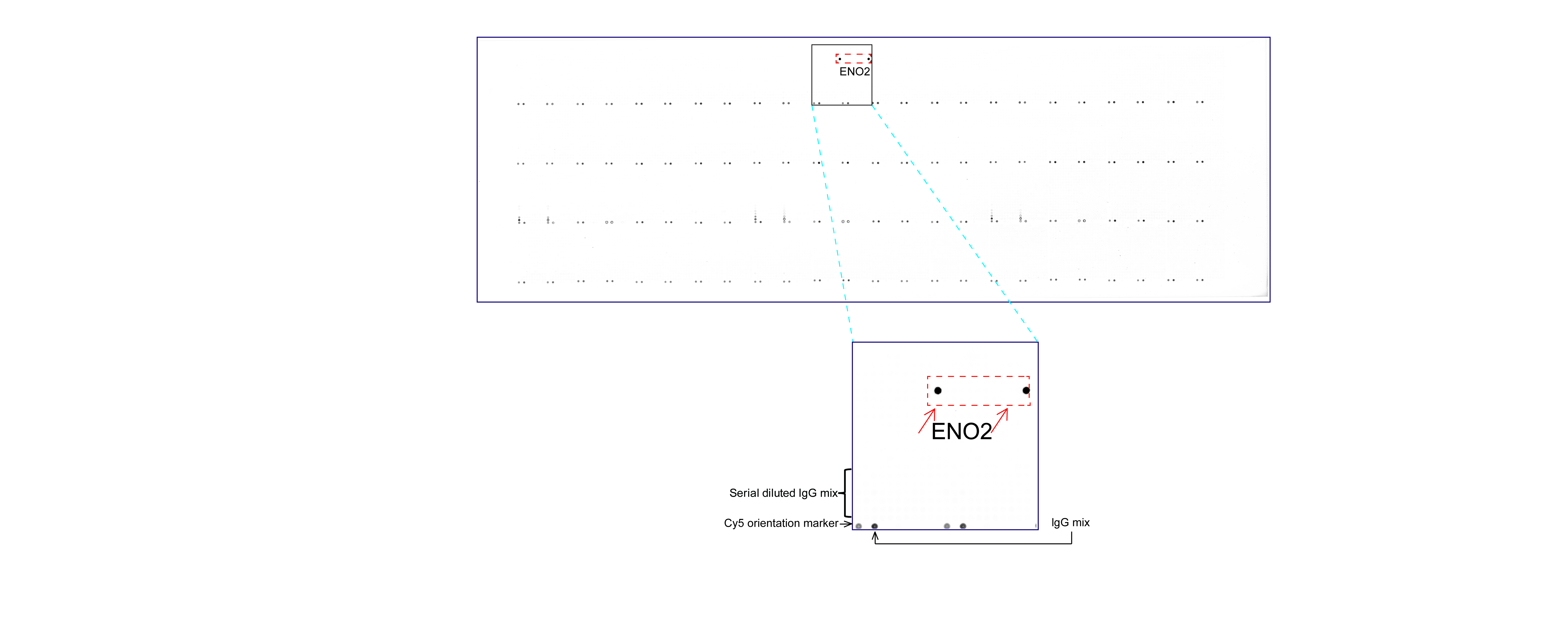
NSE is a glycolytic enzyme found in neurons and neuroendocrine cells. Early studies have shown NSE may be a good biomarker to evaluate neuron cell injury. However, NSE is also highly expressed in Erythrocytes. During hemolysis, it will be released and lead to higher level of NSE in serum, which can be a confounder. Clinical studies of NSE in mild TBI patients also show inconclusive results. High levels of NSE do not necessarily account for TBI. It can also be an indicator for tumors and stroke. Overall, NSE as a biomarker for TBI has strong limitations because of the inadequate sensitivity and poor specificity. But it can be used together with other biomarkers for the evaluation of TBI.
Products for NSE
Myelin Basic Protein (MBP)
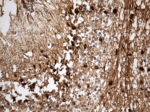
MBP is one of the most abundant proteins in myelin sheath of neurons. It is expressed by oligodendrocytes and can be released into serum after brain injury. Previous studies have shown a positive relationship between MBP serum levels and severity of brain injury in TBI patients. Higher serum MBP levels were associated with poor outcomes of the patients, which suggested a potential prognostic value of serum MBP levels in TBI. MBP is often used together with other biomarkers to predict severity and outcome of TBI. However, due to its limited sensitivity and lack of clinical validity, the popularity of MBP as a biomarker in TBI is much lower than GFAP and UCHL1.
Products for MBP

S100 Calcium-binding protein B (S100B)
S100B is a calcium binding protein that regulates calcium level in the cells. It is one of the most widely studied biomarkers for TBI. S100B is expressed in astrocytes and can be released into peripheral bloodstream after brain injury. Many studies have shown the good sensitivity of S100B testing in mild TBI (mTBI) diagnosis. Patients with normal S100B levels may not need to do the CT scan. Therefore, initial blood S100B level has a potential to be used as a screening tool to rule out mTBI. However, the specificity of S100B blood test is low. You can get many false positive results, because of other factors that can influence the S100B level in blood. In summary, S100B is a protein that can be used as a biomarker for TBI patients, but the diagnostic utility of S100B levels is limited in TBI due to its low specificity.
Products for S100B
Neurofilament Light Chain (NFL)
NFL is a neurofilament protein encoded by human NEFL gene. It is a biomarker of axonal injury for many neurological diseases such as ALS, multiple sclerosis, Alzheimer’s disease, Parkinson’s disease and Huntington’s disease. Recent studies have shown a positive correlation between NFL serum level and the progression of TBI pathology, suggesting the potential diagnostic and prognostic value of serum NFL for TBI patients. Serum NFL was better than GFAP, Tau and UCHL1 on their ability to identify TBI patients from uninjured controls. It can identify TBI patients with high accuracy even years after the injury.
Products for NEFL
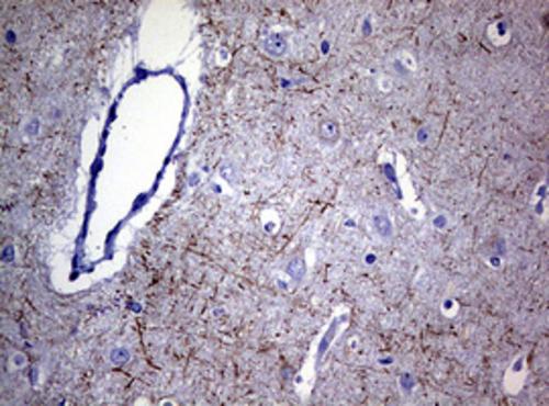
Tau protein
Tau is a microtubule associated protein (MAP) highly expressed in neurons and astrocytes. It plays an important role in microtubule stabilization and molecules transport along the microtubule. Early studies have shown a positive correlation between Tau protein and severity of brain injury in TBI patients. This relationship is further proved by various studies including a postmortem study that revealed diffuse tau pathology in patients many years after TBI. Tau pathology is one of the key mechanisms of neurodegeneration related to TBI.
Products for Tau
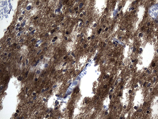
Brain-derived Neurotrophic Factor (BDNF)
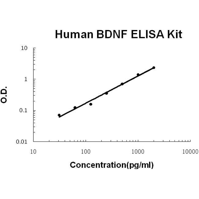
BDNF is a growth factor secreted from neuronal and glial cells, which plays an important role in neuroprotection and neuroregeneration. Because of its role in neuronal survival, BDNF can be used as a potential biomarker for TBI. Studies have shown a significant increase of BDNF mRNA level within hours after TBI (Yang et al., 1996). However, the level of BDNF declines at 24 hours, and is not significant at 36 hours post-injury (Oyesiku et al., 1999). The transient surge of BDNF following TBI suggests BDNF acts as a neuroprotective protein attempting to prevent secondary cell damage following TBI. BDNF is also a therapeutic target for TBI. Studies have shown BDNF can improve functional recovery of injured animals. However, currently there is no direct application of BDNF for TBI in experimental studies.
Products for BDNF
Antibody | Protein | Clones & Lenti | CRISPR | qPCR | RNAi | ELISA Kit


 United States
 Germany
 Japan
Japan
 United Kingdom
 China
China

